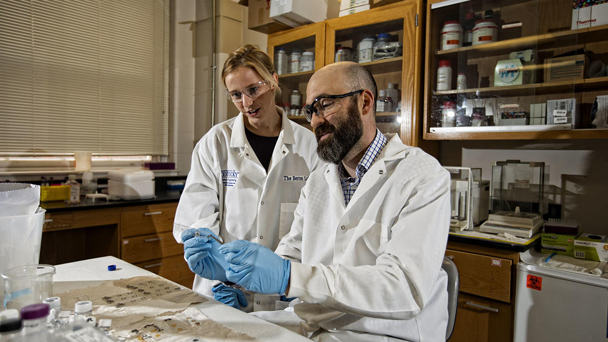
University of Kentucky Bryan Associate Professor of Chemical Engineering Brad Berron has received two grants: one from the American Heart Association and the other from the National Science Foundation. Combined, the awards total approximately $290,000.
Berron joined the faculty of the Department of Chemical and Materials Engineering at UK in 2011. He received an NSF CAREER Award in 2014.
American Heart Association: Intentional Positioning of Cells in a Decellularized Heart (abstract)
“To address the persistent shortage of patient-matched hearts, the biomedical research community has aggressively pursued the removal of cells from unmatched hearts and the repopulation of the extracellular matrix with patient derived cells. In these strategies, heart geometry is preserved, immunogenic materials are eliminated, and functional autologous cells are returned to the heart.
There are no methods to accurately control the location or density of a given cell population during recellularization, making a full reconstruction of a functioning heart impossible. To solve this problem, we will use binding motifs on the cell surface and on targeted locations in the heart scaffold to specifically load cells at their physiological location. Cell surfaces will be tagged with biotin while targeted regions in the decellularized heart will be patterned using streptavidin to achieve specific cell localization. The cells will be incubated and rinsed, yielding the functional cells in the targeted location. Additional cell types and layers are loaded sequentially. Thus, we hypothesize that targeted cell adhesion will enable both intentional positioning of multiple cell populations and higher tissue density with single-cell resolution in a decellularized heart.
Aim 1 will establish the spatial resolution, specificity of adhesion, and cell lineage generality of our novel patterned heart recellularization. Decellularized pig hearts will be biotinylated through incubation in photobiotin, exposure to patterned light, and incubation with streptavidin. Cells will be biotinylated and introduced to hearts. Pattern specificity and resolution will be determined by microscopy for a diverse array of cardiovascular cell phenotypes.
Aim 2 will examine the tissue function of patterned heart tissues. Cell viability for all phenotypes will be determined for up to one week. Cardiomyocyte, endothelial, and vascular cell function will be evaluated with immunohistochemistry and junction analysis.
The addition of autologous cells to a decellularized heart scaffold is a promising approach towards a surplus of a non-immunogenic, functioning hearts for transplantation. Current methods for cell reintroduction do not achieve the required cell density and physiological positioning of cells required for heart function. This research will discover a method for dense, physiologically-accurate assembly of heart tissue to eliminate the shortage of donor hearts.”
Assurance of Cellular Function in High-Shear Three-Dimensional Bioprinting (abstract)
“This project seeks to determine how 3D bioprinting affects cellular physiology, in particular cellular membrane transport. It is known that the high shear environment of the bioprinting nozzle impacts cellular response; however, the precise changes in the cells and whether normal function can be recovered within a range of bioprinting parameters has not been assessed. This gap in knowledge makes it difficult to determine if a 3D printed tissue actually will replicate normal (or defined pathological) physiology -- a key feature if such bioprinted tissue structures are going to be used for pre-clinical assessment of drugs or medical devices. The scientific objectives of this proposal are to: 1) quantify the magnitude and timescale of unnatural transport across the cell membrane, including multiple forms of active and passive transport, following simulated extrusion bioprinting; and 2) quantify how cellular coatings, which are hypothesized to protect cells from mechanical damage, affect the extrusion-based alterations in cellular membrane transport. In addition to measurements of membrane transport, appropriate assays for basic cell function and viability will also be conducted for each cell type. Experiments will be conducted using five cell lines (HepG2 - hepatic; Caco2 - intestinal; H9C2 - myocardial; A549 - human lung carcinoma; and mesenchymal stem cells), each of which is of current interest in the biomanufacturing arena. This collaboration will result in mutual education, with the post-doctoral fellow becoming trained in the area of regulatory affairs and the PI educating CDRH (Center for Devices and Radiologic Health) team members at the FDA on emerging technology for encapsulated cell systems and devices.”
